By Dr. Robert L. Bard
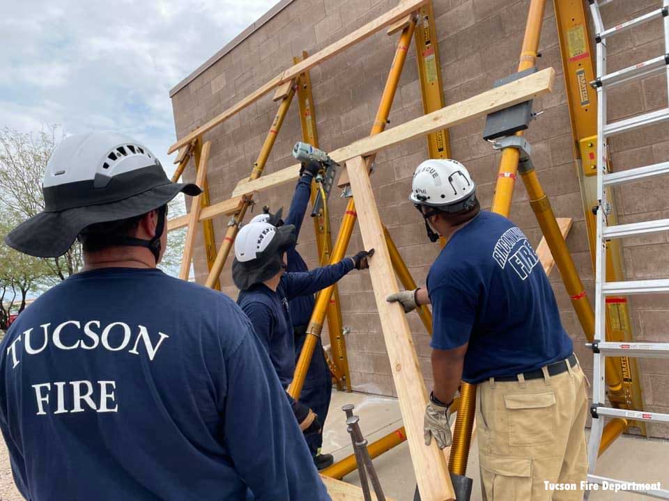
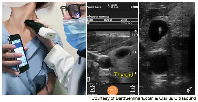
Portable handheld sonogram units have been used by the militaries of advanced nations for years and have quickly identified all levels of trauma from a simple bruise to a serious medical issue, and continuing advancements in portable medical diagnostic technologies are now helping rescuer in their work. Blunt trauma to the rib cage could range in severity from a hematoma (internal bleeding), a fracture or a penetrating lung contusion. Today’s compact ultrasound systems are wireless and WIFI-powered, allowing scans to be sent to any area of the world for medical overreading in real time.
With respect to victims trapped by settling wreckage, tissue damage is diagnosable and the site for amputation may be demarcated so appropriate triage and definitive treatment may be planned expediently. Advanced military targeting cybernetics that allowed Israeli jets to selectively pinpoint a terrorist cell in the middle of an 11-story high rise complex was used to relocate the probable escape routes of the potential survivors during the recent building collapse in Surfside, Florida. It also helped identify their possible placement within the computerized trajectory of human and structural movement, allowing one of the rescue workers to find the body of his missing daughter intact.
Point of Care Response: When Time Is of the Essence
It is important to note that up to 90 percent of rib fractures are missed by standard X-ray views because the bone is a rounded structure with a curved outline so the break of the boney outer cortex must be X-rayed at an exact tangential view. In the case of the sonogram, targeting the injury results in a higher rate of success as the patient drives the probe over the area of greatest pain.
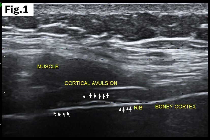
(1) Rib cortical avulsion: This type of fracture is painful, produces shortness of breath but does not lead to a punctured lung surface and may be treated conservatively.
A hematoma shows up as an oval mass of fluid, appearing as black on the white background screen. A pulmonary contusion produces thickening of the outside of the lung (pleural surface) and the extent of the lung damage is measurable. At this point, use of a pulse oximeter verifies the oxygenation of the blood by documenting the pulse rate. In the case of a true lung puncture (pneumothorax), the patient is usually unstable and needs emergency care. A partial or complete pneumothorax is diagnosed as complete absence of the sliding motion of the pleural surface lining.
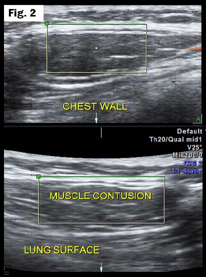
(2) Chestwall contusion: Soft tissue swelling of the muscle demonstrates intact pleural surface without pneumothorax.
A sprained ankle, common in debris excavations, is readily amenable to portable ultrasound imaging. Bone, ligaments, tendons, and muscle are clearly visible on the image and the diagnosis of a sprain is verified as the patient places the probe over the site of maximal tenderness. Since first responders are often mentally engaged in the recovery and may be ignoring the pain, the occurrence of tendinitis is also available by sonography. Inflamed tissue needs blood vessels to heal, and the Doppler blood flow aspect of the unit will reveal the number of vessels that are supplying the injury with oxygen. Blood flow is red in color on the system, and this is another factor in assessing the injury and following progress of the treatment. No viable arterial flow is noted in the case of traumatized, dead tissue and the viable muscle is differentiated from the gangrenous limb.
Burns
Whereas a burn on the skin is obvious, the depth is not observable, and the tissue viability is unknown. First-degree burns show skin thickening that generally corresponds to the red area. Second degree burns demonstrate greater enlargement of the dermis and often blister fluid, which further decreases the clinicians view of the extent. Doppler blood flow is present in both cases of first- and second-degree thermal damage. Third-degree burns show no blood flow and the dead tissue images black as contrasted to the first- and second- degree regions. As healing progresses, the thickness of the skin lessens as does the healing blood flow. This process is quantifiable by 3-D Doppler histogram analysis providing a percentage (%) number of the capillaries in the site. More importantly, for skin grafting procedures, the critical issue of tissue health and viability is measurable with this noncontact, noninvasive modality.
Pulmonary Emboli
Deeper damage to the muscle, fascia, tendons, and vascular structures may necessitate the requirement for anticoagulation to prevent blood clots from travelling to the lungs (pulmonary emboli). Ultrasound also analyses the cardiac and pulmonary effect of emboli to the lung vasculature. This entity also affects the adjacent cardiac structures which are investigated at the same time as the chest is studied by sonographic technologies.
Response to Occupational Exposures
Anyone who received a letter from the Department of Health alerting them to the high mercury, arsenic, or lead level in a recent blood or urine test knows that this is a wakeup call to further evaluate the problem. However, most of the state health standards were based on toxicity to the unborn fetus in pregnant mothers. This is usually not applicable to most first responders. For some reason, the early blood test abnormalities do not always match the results of the 24-hour urine collection, which is supposedly more accurate. Many physicians today accept a high blood mercury level as a sign of health from greater consumption of fish.
Another unknown is the combination of toxins with trauma and particulate matter at high rise disasters. Many cancers and chronic diseases do not manifest fully until months, years, or even decades after the initial body insult. This is clearly identified from the reports of cancer cases from the 1975 NY Telephone Exchange Fire and the cancer registry for the 9/11 attack aftermath, which is still revealing a significant incidence of skin, lung and prostate cancers nearly 20 years later.
Medical researchers and prevention advocates in rescue units often reference medical data from landmark cases (like 9/11) for their recorded patterns of illness. Quantifying the percentage of job-related exposures, the most common tumors recorded after 9/11 are basal cell skin cancer, squamous cell skin cancer, and malignant melanoma, then prostate, breast, thyroid, lung, and kidney [1]. Other statistics from the Firefighter Cancer Support Network* have noted the incidence as prostate (13.7 percent), skin (8.4 percent), colon (7.1 percent), bladder (6.9 percent), and testicular (5.5 percent) [2].
Modern diagnostic imaging allows non biopsy analysis of all three skin cancers and prostate tumors. Blood-flow Doppler measurements predict which tumors are aggressive and which may be watched. Lung cancers are best tracked by CT and PET/CT isotope imaging. The NY Cancer Society stated in the 1990s that 3 percent of prostate cancer is truly lethal (bladder cancer is 40-50-percent deadly by comparison), meaning that low grade prostate nodule may be assessed on a yearly basis for change in size and vascular data for malignant potential. The U.S. government agrees that men should submit to PSA testing only after discussion with their physicians. Ultrasound and laser-MRI guided focal treatments permit men to handle the problem with outpatient, minimally invasive procedures.
Utilities: Changes and Challenges for the Fire Service
Toxins from Plastics
Modern building materials and appliances ignite and off-gas more aggressively, setting off a range of toxic compounds. At high temperatures, the chemical bond of these compounds weakens and breaks down as the higher energy state shifts to a more natural and stable lower energy scale in accordance with the laws of thermodynamics. As plastics slowly disintegrate, chemical bond breakage releases the energy as heat and other byproducts such as gases and particulate matter. Eventually, the buildup of these offshoots of energy transfer sufficiently increases the local heat envelope and the halo of combustible gases and matter, which ignite into flame or causes explosive outbursts. Unfortunately for the unsuspecting in the fire zone, the toxic fumes often contain carbon monoxide, which deprives the body of oxygenated hemoglobin, inducing weakness, confusion, and instability. This poisonous, invisible cloud results in most deaths during mass-casualty thermal disasters. In many conflagrations of large structures, most victims were lifeless before being engulfed by the flames. Spontaneous combustion events occurred at the Surfside condo site days after the collapse further hindering rescue efforts.
Toxic Emissions
Plastics are formed from the distillation of oil products turn into chemicals; typically, new synthetics such as plastic, glues and pesticides contain helping additives such as fire retardant and plasticizers. Decomposition to a more stable chemical state may be slow (years), producing irritation, or rapid, causing fire and explosion. In building scenarios, we deal with high-temperature oxidative pyrolysis with carbon monoxide fumes, the acidic gases emanating in the pre-flame stage and damage of the tissues by direct chemical burns, accumulation of fluids into the tissue spaces, and hemorrhage from the inflamed blood vessels. Emissions from burning plastic such as polyvinylchloride (PVC), PCB, urethane foam, plumbing pipe, and polystyrene include carbon monoxide, benzene, hydrogen chloride, and hydrogen cyanide gas. These substances are not only acutely toxic to organ systems but are known carcinogens. The “salty” reference links to the hazardous soot that is inhaled, ingested (swallowed), and physically in contact with the skin, hair, and sweat glands. While these chemicals are immediate asphyxiants and damage organs that need high levels of oxygenated blood such as the heart, brain, liver, and kidney, the long-term carcinogenic effects have been systematically categorized and extensively studied from the September 11, 2001 cancer registry.
Toxins and Cancer
Major illnesses from inhaling smoke include loss of immunity to respiratory tract infection, airway sensitization (chronic cough), lung damage, sinus trouble, problems with peripheral circulation, and heart and neurological/psychological disorders. The most common cancers attributed to the New York Telephone fire in 1975 were in the larynx, liver, skin, throat, and brain, and were detected through the years 1980 to the present. The remaining survivors are still developing cancers to this day.
Risk Assessment: ‘Listen’ to Your Skin
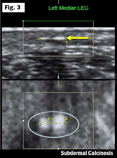
The skin is the largest organ in the body and, like the liver and lungs, is a filter for toxins. Ultrasound advanced technologies have assessed lung and liver damage accurately for the past few years. [Bard] High-resolution ultrasound now is used to interrogate the skin for signs of heavy metal toxicity. This is a natural area of investigation since the toxins from an incendiary event remain on the skin and the adjacent protective gear. We know that some inflammatory changes in living tissue heal by scar formation, which may calcify as submillimeter foci that are observable by the micro-ultrasound units. Scar tissue has been extensively studied with sonograms for depth and quality as well as the effect on adjacent structures.
(3) White dots of heavy metal poisoning in dermis. The light gray dermis expands from 1 to 2 mm thickness and darkens, indicating inflammation. Arrows point to “dots” of uranium deposition in the 4 mm thickened dermal layer.
Calcium deposits are proportional to the tissue damage and may be a measure of toxic exposure. This has been documented for heavy metal poisoning and radioactive substances found in well water such as uranium and radon. The dermis provides a mirror into the body that may be noninvasively imaged in minutes to serve as a barometer for exposure and, by extension, cancer risk. The dermal microcalcifications are so small that they are rarely visualized by X-ray or CT scans. 3D volumetric ultrasound groups these “white dots” and counts the number in the captured tissue volume. When the bright areas are displayed on the screen, they appear like twinkling stars as in the painting by Van Gogh “Starry Night.” It’s often so obvious that the patients are compelled to comment on the visual drama that lies within their living skin. This serves as a baseline for treatment follow up as well as an alert to get checked for cancer development in other organs. A common treatment for heavy metal poisoning is “chelation,” which often reduces the toxic burden and improves symptoms. Quantitative verification of treatment success is available through serial sonogram calculation of the white dot density.
REFERENCES
CDC/WTC Data: https://www.cdc.gov/wtc/ataglance.html#top15Cancers
FCSN Web site: What research supports the link between firefighting and cancer? https://firefightercancersupport.org/resources/faq/#:~:text=The%20top%20cancers%20for%20male,and%20testicular%20(5.5%20percent)
World Trade Center (WTC) Health Program Statistics
Bard, Robert L., Dynamic Contrast-Enhanced MRI Atlas of Prostate Cancer, Springer, 2009.
Bard, Robert L., Fütterer, Jurgen J., Sperling, Dan, Image Guided Prostate Cancer Treatments, Springer, 2014.
Bard, Robert L., Image Guided Dermatologic Treatments, Springer, 2019.
Bard, Robert L., Image-Guided Management of COVID-19 Lung Disease, Springer, 2021.
ROBERT L. BARD, MD, PC, DABR, FASLMS, is recognized for his specialized work in advanced cancer diagnostic imaging. He co-founded the 9/11 CancerScan program to bring additional diagnostic support to all first responders from Ground Zero. His main practice in midtown, NYC (Bard Diagnostic Imaging, www.CancerScan.com) uses the latest in digital imaging technology and has been also used to help guide biopsies and, in many cases, even replicate much of the same reports of a clinical invasive biopsy. Imaging solutions such as high-powered sonograms, Power Doppler Histogram, sonofluoroscopy, 3-D/4-D image reconstruction, and the Power Doppler Histogram are safe, noninvasive, and do not use ionizing radiation. It is used as a complement to find anomalies and help diagnose the causes of pain, swelling, and infection in the body’s internal organs while allowing the diagnostician the ability to zoom and ‘travel’ deep into the body for maximum exploration.